+7 (929) 727 53 60 Травматология / Ортопедия
Comparative spectral analysis of the component composition of bioimplants making in different ways for the treatment of gingival recession
P.E. Timchenko1, E.V. Timchenko*1, L.T. Volova2, O.O. Frolov1, V.D. Meseyarakov1, Pugachov E.I.2
1 Samara National Research University, 34, Moskovskoye shosse, Samara, 443086, Russia
2 Samara State Medical University, 89 Chapayevskaya St., Samara, 443099, Russia


The spectral study results and comparative evaluation of the component composition of the surfaces of hard palate implant samples made by the technology "Lioplast", used in the clinic in the field of atrophic processes in multiple recessions of the gums, using the method of Raman spectroscopy (RS) are presented. It is shown that Raman spectroscopy can be used to assess changes in the composition of implants based on the hard palate during their processing. The coefficients were introduced and a two-dimensional analysis was carried out, which showed that the main components are preserved and DNA/RNA is removed during processing, which improves the quality of the material that provides the possibility of a good clinical effect in the treatment of multiple recessions of the gums. Keywords: Raman spectroscopy, spectral analysis, bioimplants, gingival recession, “Lioplast”.
The recovering of atrophied gingival tissues due to adentia is a very actual problem. There is also a risk of immune implant rejection and an increase in the survival time of the recipient during the transplantation of native implants [1]. Therefore, the main problem of tissue engineering is the development and quality control of materials that can restore or replace a particular function of damaged tissues and organs. In this disease, patients are usually concerned about aesthetics, increased sensitivity of the exposed roots of the teeth, unpleasant sensations when cleaning them, caries [2]. The most significant factors contributing to the development and progression of the recessions are: inflammatory processes in the oral cavity caused by poor hygiene, age-related physiological changes, heredity [3]. Allogeneic biotissue is proposed to use for the first time in the world practice in the treatment of this pathology as a plastic material. The allogeneic biotissue made by the original domestic technology "Lioplast" ®" and based on the hard palate of man. The use of allogeneic material, in this case, is the most advantageous solution, since it can be used in multiple recessions. A successful outcome of such operations depends on the quality and technologies of production materials, maintaining the desired biological substances such as collagen, glycosaminoglycan, proteoglycans [4] and the removal of cellular components (DNA, RNA). There are many methods to control the quality of treatment of bioimplants, including histological, molecular and biochemical analysis. These methods make it possible to quantitatively assess the structural composition of biomatrix, but this analysis is destructive and it takes a significant amount of time to display the results of histological and cytological analysis. Therefore, to assess the composition of biological tissues it is necessary to use optical control methods, which are non-invasive. Raman spectroscopy is one of the most common optical methods for assessing the quality of a bioimplant [5]. The Raman method allows to control the component composition of biomaterials, and has a number of advantages, such as ease of sample preparation, minimally invasive, a large amount of information obtained, the efficiency of the study [4, 5, 6]. The aim of this work is determining the most important criteria for assessing the component composition of solid palate samples at the manufacturing stages using the non-destructive Raman spectroscopy method.
MATERIALS AND METHODS RESEARCH
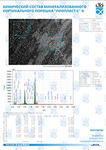
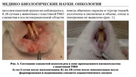
Samples of bioimplants of the lyophilized palate were used as objects of research. The studied samples were processed using the technology "Lioplast"® (TU-9398-001-01963143-2004). Component analysis of bioimplants from hard palate of lyophilized and lyophilized samples was carried out. As the main method of analysis of lyophilized palate samples was used the method of Raman spectroscopy, implemented by high-resolution digital spectrometer Shamrock sr-303i (figure 1), combined with the laser module LuxxMaster LML785.0RB-04 (power up to 500 mW, wavelength 785 nm) and built-in cooled camera DV420A-OE, providing a spectral resolution of 0.15 nm (spectral range 200-1200 nm). Selection of the Raman spectrum against the background of autofluorescence was carried out by polynomial approximation of fluorescence and its subtraction from the recorded spectra in Wolfram Mathematica. The approximating line (fifth degree polynomial) of the autofluorescence component was determined on the selected interval 400-2200 cm-1 using an iterative algorithm and then this component was subtracted to obtain the selected Raman spectrum [7]. The analyzed spectra in the processing cleansed from noise smoothing by a median filter of 7 pixels.
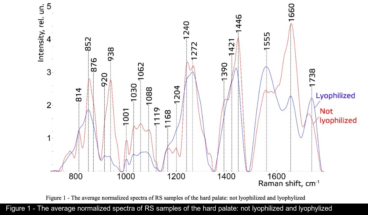


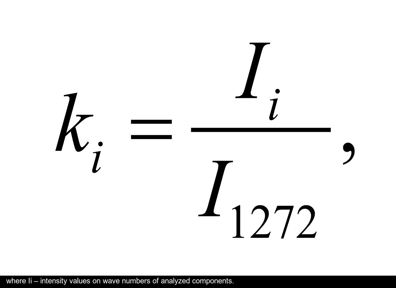
We have introduced relative coefficients for a relative quantitative assessment of the component composition of solid palate samples. A relatively constant component in the studied samples was amide III [4, 10] (figure 1), corresponding to the region of wave numbers 1230-1280 cm-1 , so the amplitude of the divided line of amide III was used as a denominator (I1272) in the introduced coefficients (k):
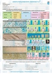
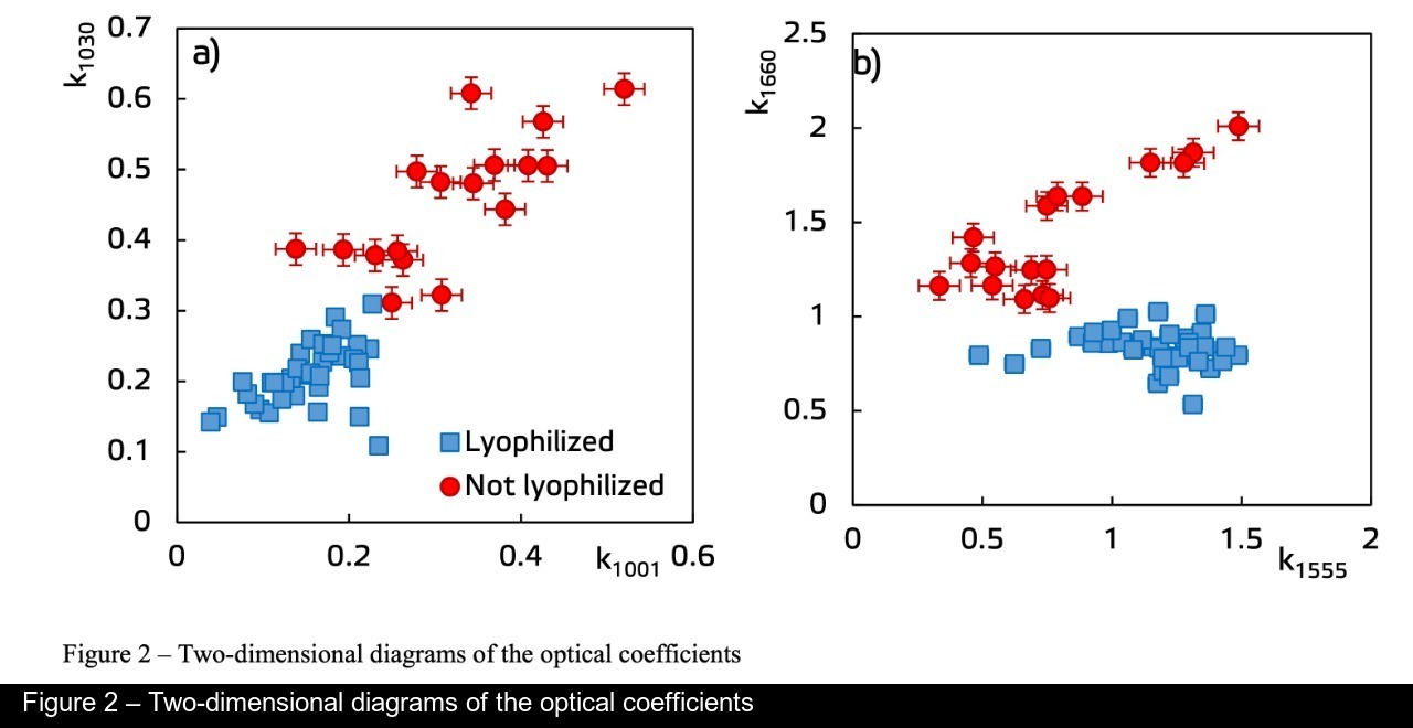
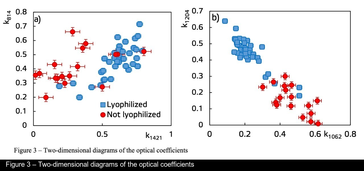
To assess the quantitative content of the main components of bioimplants, a two-dimensional analysis of the optical coefficients was performed. It allows to compare the quality of the samples. A detailed analysis is given in figures 2-3, which presents two-dimensional diagrams of the introduced coefficients and also shows the differences and similarities between the two groups of samples.
Figure 2a shows the values of the optical coefficients k1001 and k1030, reflecting the relative concentration of phenylalanine in collagen and proteins included in the biomatrix. Taking into account the data in figure 1, it can be said that there are significant differences between the two groups, i.e. the relative concentration of phenylalanine after lyophilization decreases. The relative concentration of amide II is higher in lyophilized samples (k1555), and the k1660 coefficient reflecting the relative concentration of amide I is higher for untreated samples (figure 2b). From figure 3a it is also seen that the values of the coefficients k814 and k1421, reflecting the relative concentration of DNA / RNA in the lyophilization process remain virtually unchanged, suggesting the impossibility of a complete withdrawal of the remnants of DNA / RNA from the sample. The values of the coefficients reflecting the relative concentrations of glycosaminglicans and tyrosine, I1062/I1272 and I1204/I1660 vary (0 < I1062/I1272 < 0.3; 0.3 < I1204/I1272 < 0.7) for lyophilized samples, and (0.35 < I1062/I1272 < 0.6; 0 < I1204/I1272 < 0.3) without special treatment using the "Lioplast" technology (figure 3b), that reflects a drop in the relative concentration of gags relative to collagen structures (amide III) in the manufacturing process.
The comparative spectral estimation of the component composition of the surfaces of the samples of hard palate implants made by the "Lioplast" technology with and without lyophilization was carried out. It was found that the main differences are manifested in the wave numbers 1002 and 1030 cm-1 (phenylalanine), 1062 cm1 (GAGs), 1204 cm-1 (tyrosine), 1421 and 814 cm-1 (DNA / RNA), 1555 and 1660 cm-1 (amide II and amide I). The coefficients were introduced and a two-dimensional analysis was carried out, which showed that after lyophilization treatment the main components of biomatrix are preserved, which increases the quality of the material providing the possibility of a good clinical effect in regenerative processes. It is shown that after the treatment of bioimplants by lyophilization, the components that negatively affect their quality are removed. While the necessary level of extracellular matrix is maintained: glycosaminglicans, collagen, proline, hydroxyproline and phenylalanine, which play an important role in the process of implant engraftment, and obtaining a high-quality extracellular matrix.
The studies were carried out with financial support of Russian Foundation of Basic Research (RFBR), project 18-32-00004.
[1] Muslimov, S.A., "Morphological aspects of regenerative surgery," Ufa, Bashkortostan 168 (2000).
[2] Johnson, J. F., "Modern practice in crown and bridge prosthodontics," OperativeDentistry 184–186 (1971).
[3] Ganja, I. R., "Gum recession. Diagnosis and treatment methods," Samara: «Sodruzhestvo» 52-54 (2007).
[4] Chen, H., "In vivo spinal nerve sensing in MISS using Raman spectroscopy," In Proceedings of SPIE 9802(98021), (2016).
[5] Saxena, T., "Raman spectroscopic investigation of spinal cord injury in a rat model," Journal of Biomedical Optics 16(2), 27003 (2011).
[6] Timchenko, P. E., "Application of Raman spectroscopy to assess the condition of bone and cartilaginous biopsy specimens," Journal of Optical Technology 84(6), 423-425 (2017).
[7] Zhao, J., "Society for applied spectroscopy," 61(11), 1225–1232 (2007).
[8] Timchenko, P. E., "Optical Analysis of Implants from the Dura Mater," Optical Memory and Neural Networks 27(1), 46–52 (2018).
[9] Timchenko, P. E., "Diagnostics of bone implantat and control of their process osteointegration with of a method confocal microscopy," Computer Optics 35(2), 183-187 (2011).
[10]Timchenko, P. E., "Experimental studies of hydroxyapatite by Raman spectroscopy," Journal of optical technology 85(3), 130-135 (2018).
-
Research of component composition of mineralized bone implants by Raman spectroscopy
 ПодробнееФайл статьи P. E. Timchenko, El. V. Timchenko, L. T. Volova, O. O. Frolov, V. D. Meseyarakov, E. I. Pugachov, "Optical estimation of the composition of bone implants during processing," Proc. SPIE 11074
 -
Raman spectroscopy for evaluation of dura mater based grafts
 Файл статьи P. Е Timchenko, Е. V Timchenko, L. Т Volova, О. О Frolov, А. U Kulabuhova, N. K KiykoПодробнее
 -
Analysis of the mineral component for cortical bone tissue by Raman spectroscopy after ovariectomy and its treatment with allogeneic hydroxyapatite
 Файл статьи E. Timchenko1, P. Timchenko1, E. Pisareva1, M. Vlasov2, L. Volova2, I. Fedorova1, A. Tumchenkova1, M. Gorchenkova1 and A. Subatovich1Подробнее
 -
Research of component composition of bioimplants for treatment of gum recession using a Raman spectroscopy method
 Файл статьи P. Е. Timchenko1, Е. V. Timchenko1, L. Т. Volova2, О. О. Frolov1 and E. F. Yagofarova1Подробнее