+7 (929) 727 53 60 Травматология / Ортопедия
Chemometric analysis of bioimplants of bone tissues during their manufacture
CHEMOMETRIC ANALYSIS OF BIOIMPLANTS OF BONE TISSUES DURING THEIR MANUFACTURE
P.E. Timchenkoa, E.V. Timchenko*a , L.T. Volovab, O.O. Frolova
aSamara National Research University, 34, Moskovskoye shosse, Samara, 443086, Russia
bSamara State Medical University, 89 Chapayevskaya St., Samara, 443099, Russia
ABSTRACT
The results of expanded spectral analysis of donor bone implants using the Raman spectroscopy method are presented in the work. Mathematical methods of improving resolution of spectral contours and chemometric analysis are used for evaluation of component composition of the implants. It is shown that the Raman spectroscopy can be used for evaluation of relative concentration of mineral and organic components in extracellular matrix of bone tissue. Spectral features of the samples of bone tissues demineralized and processed with ultrasound with different degree are found as a result of the study. The criteria allowing evaluating the component composition during manufacture of bioimplants are suggested in the work.
Keywords: Raman spectroscopy, spectral analysis, bone bioimplants, chemometrics, demineralization, “Lioplast”.
Downloaded From: https://www.spiedigitallibrary.org/conference-proceedings-of-spie on 14 May 2020 Terms of Use: https://www.spiedigitallibrary.org/terms-of-use Tissue Optics and Photonics, edited by Valery V. Tuchin, Walter C. P. M. Blondel, Zeev Zalevsky, Proc. of SPIE Vol. 11363, 113632A · © 2020 SPIE CCC code: 0277-786X/20/$21 · doi: 10.1117/12.2565723 Proc. of SPIE Vol. 11363 113632A-1


The results of expanded spectral analysis of donor bone implants using the Raman spectroscopy method are presented in the work. Mathematical methods of improving resolution of spectral contours and chemometric analysis are used for evaluation of component composition of the implants. It is shown that the Raman spectroscopy can be used for evaluation of relative concentration of mineral and organic components in extracellular matrix of bone tissue. Spectral features of the samples of bone tissues demineralized and processed with ultrasound with different degree are found as a result of the study. The criteria allowing evaluating the component composition during manufacture of bioimplants are suggested in the work.
Keywords: Raman spectroscopy, spectral analysis, bone bioimplants, chemometrics, demineralization, “Lioplast”.
Ensuring full regeneration of bone tissues in a damaged area of a bone is one of the acutest problems of modern medicine, despite the accumulated knowledge in this field. It can be solved by creating optimal conditions for regenerating processes in the areas of its resorption. One of the ways is using bone plasty materials [1, 2]. Among them are allogeneic implants made of human tissues that are optimal materials for reconstructing the damaged musculoskeletal system. The use of such implants does not disrupt homeostasis and metabolism of connective tissues and function of life support system of recipient, unlike autoplasty and xenoplasty and using synthetic materials [3]. After special processing allogeneic materials almost completely lose their antigenicity and when put into the body do not have a negative effect on it. They serve as a matrix, a conductor, gradually fully dissolves, and in their place new bone tissue is formed [4].
The success of such surgeries depends on their quality and the technology of bioimplant manufacture, aimed to maintain the necessary biological substances, involved in regenerative process, as hydroxyapatite, collagen, glycosaminoglycans [5], and to remove the cellular components (DNA, RNA) – the main factor of antigenicity.
The process of bioimplant manufacture requires constant monitoring of its quality and evaluating the organic compound. The quality of bioimplants can currently be assessed in vitro using a series of morphological, morphometrical, biochemical studies, but their disadvantage is the long process of information receiving and the destructive impact on a subject. Therefore the use of optical methods is rather promising as they can be used as screening tests, they are performed rapidly, low-cost and do not destroy the presented samples [6, 7].
Among the physical methods the widely used for control of the implants made of bone tissues are scanning electron microscopy [8], X-ray spectroscopy [9] and the Raman spectroscopy method [10, 11], that has certain advantages and allows making real time nondestructive qualitative and quantitative analysis of composition of biological objects and provides information about molecular structure with high spatial resolution.
The method of Raman spectroscopy is used in the work [12] for studying the crystal structure of human hard dental tissues in the dental caries process. The spectral features of samples of hard dental tissues in different pathological processes were found as a result of the experimental study. The authors also concluded that the method is informative for studying the composition and structural features of biomaterials.
The aim of this work: evaluation of mineralized bone implants during the process of their manufacture using the method of Raman spectroscopy and the methods, making the spectra more informative.
MATERIALS AND METHODS OF RESEARCH
Raman spectroscopy method, implemented by the experimental stand, including the Raman probe RPB-785 (focal length of 7.5 mm), combined with the laser module LuxxMaster LML-785.0RB-04 (power up to 500 mW, wavelength of 784.7 ± 0,05 nm) and the high-resolution digital spectrometer Shamrock sr-303i, providing spectral resolution of 0.15 nm, with the build in cooling camera DV420A-OE (spectral range of 200-1200 nm), was used as the main method of analysis of bioimplants.
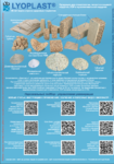
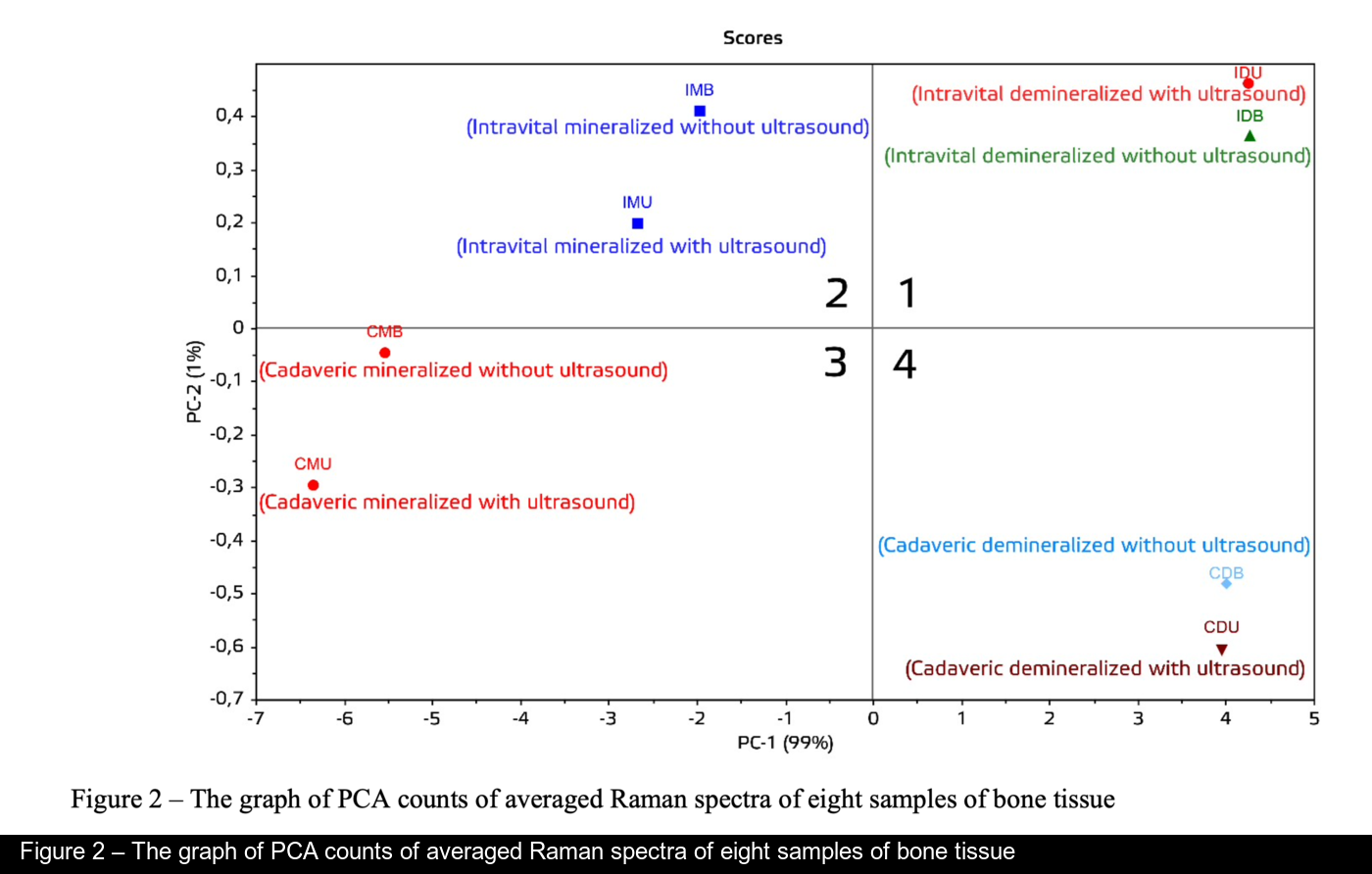
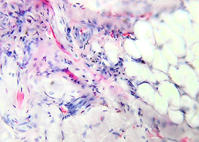
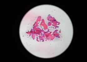
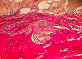
The subjects of the study were 48 samples of cancellous bone bioimplants in the form of a cube with sides measuring 5*5*5 mm, made using "Lioplast"® technology (technical specifications TU-9398-001-01963143-2004). All samples were divided in two groups according to the way of their manufacture: allogenic (cadaverous) and intra-operatively resected. The samples in every group were divided in four subgroups according to the amount of demineralization and ultrasound processing (eight groups of samples total). The spectra of each of six sides of sample were taken in different points and averaged.
The technological process of bone bioimplant manufacture included low frequency ultrasound processing of tissue (24- 40 kHz for 2-3 minutes), that ensured maximum spongious degreasing and removal of stroma and bone marrow cells from trabecular area of the bone. Demineralization of the bone tissue samples was made using mild solution of hydrochloric acid. The final stage of processing included bone lyophilisation using ALPHA 2-4 LSC device, which is freeze-drying the samples. Then the sealed material was finally sterilized using radiation techniques.
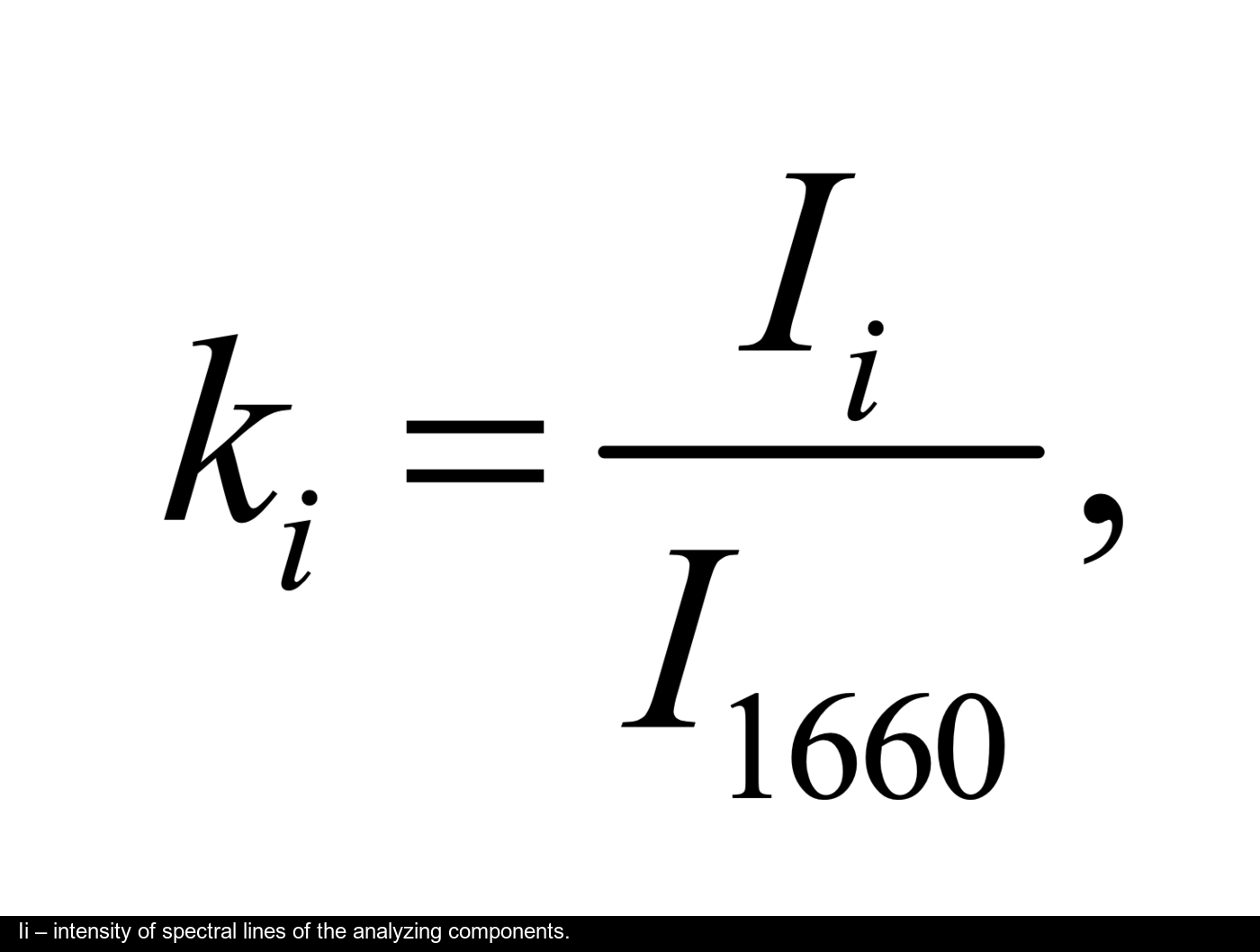
The spectra processing was made in the software Wolfram Mathematica 10 and included clearing up noises by the smoothing median filter (7 points). Then in the chosen area of 300-2200 cm-1 approximating line (an eighth order polynomial) of autofluorescent component was determined using iteration algorithm [13] and then this component was subtracted receiving allocated Raman spectrum. The error of the used coefficients did not exceed 4% [11].
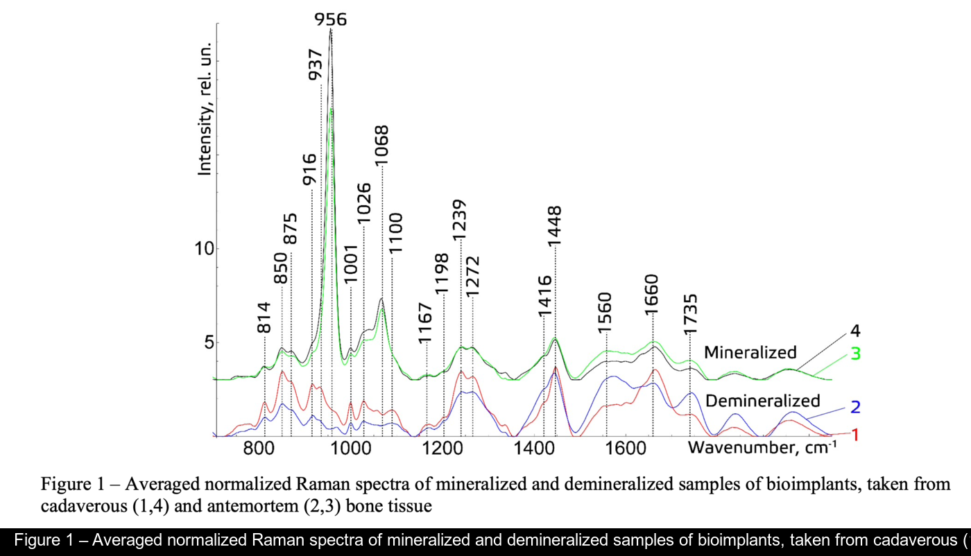


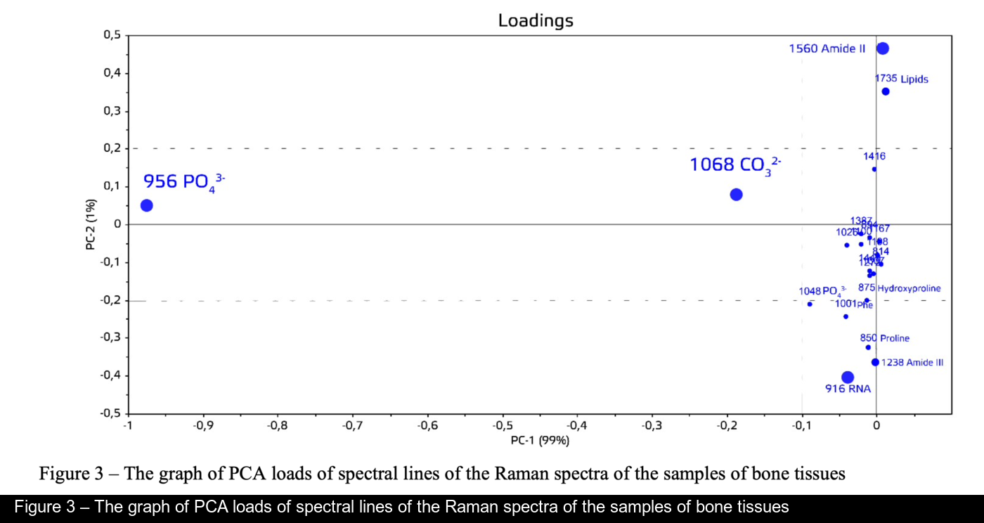

Figure 1 shows the distinctive Raman spectra of mineralized and demineralized samples, obtained from different sources in the area of 750 – 2000 cm . The main differences are seen in the Raman lines of 814, 850, 956, 1001, 1026, 1068, -1 1239,1272,1560and1735cm-1.
The main criteria of evaluation of component composition of mineralized bone grafts made using "Lioplast" technology during their manufacture were established.
The introduced coefficients allow evaluating the degree of influence of ultrasound processing, demineralization and the source of bone tissue.
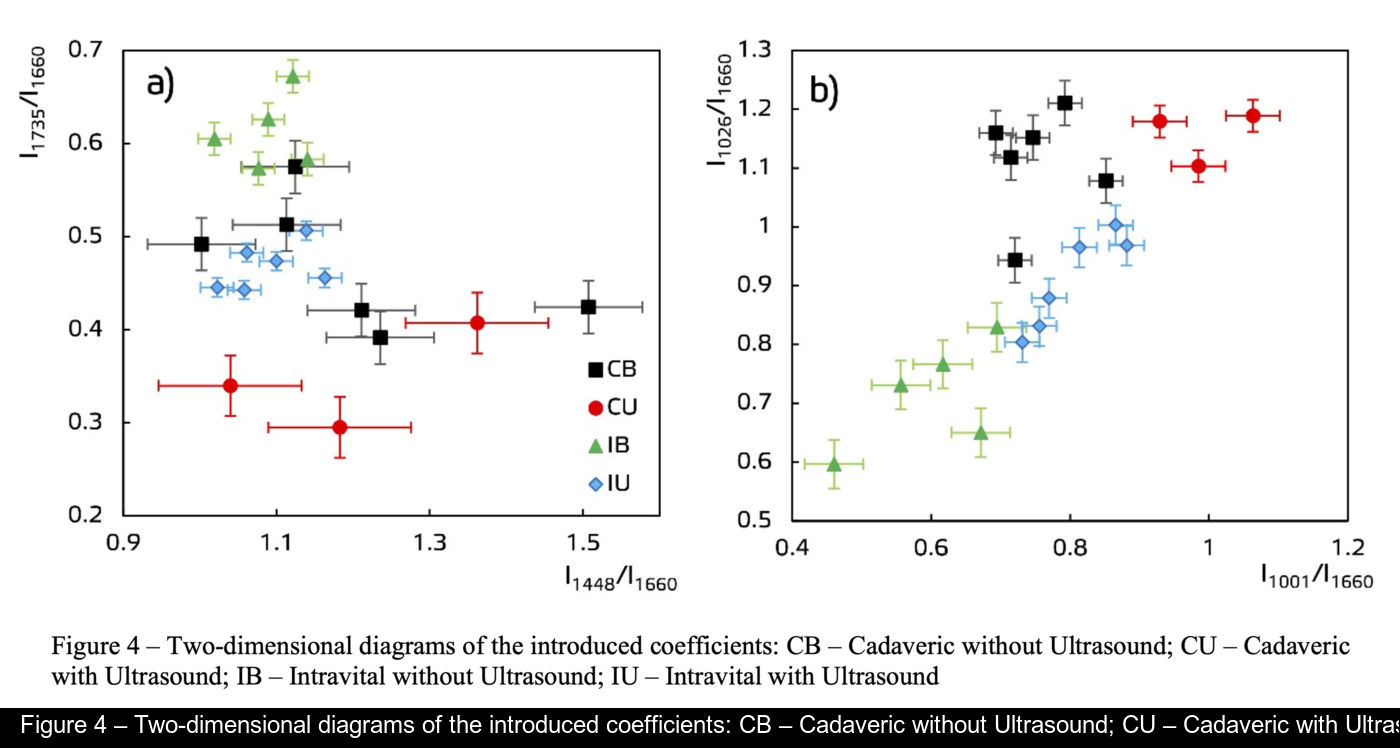
It was established that the main differences are seen in the Raman lines of 1448, 1735 (lipids & fatty acid), 850 and 875 cm-1 (proline & hydroxyproline), 1001 and 1026 cm-1 (phenylalanine) and 1272, 1560 cm-1 (amide II, amide III).
It was shown that optical method of bioimplant assessment by the introduced spectral ratio can be further used for optimization of the process of its manufacture due to improvement of quality of the manufactured material and selection of individual parameters of its processing.
The research was carried out with support of Russian Foundation of Basic Research, project 18-315-20017/18
-
[1] Muslimov S.А. Morphological Aspects of Regenerative Surgery // Ufa.: Bashkortostan. 2000. 168 p.
-
[2] Lekishvili М.V. Technologies for manufacturing bone plastic material for use in reconstructive surgery //
dissertation of Doctor of Medical Sciences: 14.00.41, 14.00.22 Moscow. 2005. 47 p.
-
[3] Saveliev V.I., Kornilov N.V., Ivankin D.Е., Linnik S.А. Allotransplantation of formalinized bone tissue in
traumatology and orthopedics // Saint Petersburg.: MORSARAB. 2001. 208 p.
-
[4] Ladonin S.V., Belozertseva Е.А. Application of allogeneic demineralized bone implants in the treatment of
chronic osteomyelitis in an experiment // Topical issues of tissue and cell transplantology: Moscow, CITO. 2007. 27 p.
-
-
[5] Chen H., Xu P.W., Broderick N. In vivo spinal nerve sensing in MISS using Raman spectroscopy // In Proceedings of SPIE, Las Vegas: Society of Photo-optical Instrumentation Engineers (SPIE). 2016. V.9802. pp. 98021L.
-
[6] Timchenko P.E., Timchenko E.V., Volova L.T. etc. Raman spectroscopy method for the evaluation of bone bioimplants made using the »lyoplast» technology from cadaveric and in vivo resected bone tissue // Journal of Physics: Conference Series. — 2018. — Vol. 1038. Issue 1.
-
[7] Timchenko P.E., Timchenko E.V., Volova L.T., Frolov O.O. Spectral Analysis of Organic Components of Demineralized Bone Biografts // Optics and Spectroscopy (English translation of Optika i Spektroskopiya) 2019. — Vol. 126. Issue 6. — P. 769-775.
-
[8] Kolosov V.Y. Studying nanomaterials using the methods of scanning electron microscopy: methodological gidelines // Ural State University. Ekaterinburg. 2008. P. 17
-
[9] Ungureanu D.N. Thermal stability of chemically precipitated hydroxyapatite nanopowders // International journal of biology and biomedical engineering. 2011. V.5. I.2. P. 57-64.
[10]Koljenovic S., Schut T.B., Vincent A., Kros J.M., Puppels G.J. Detection of meningioma in dura mater by Raman spectroscopy // Analytical Chemistry. 2005. V.77. 7958–65.
[11] E. V. Timchenko ; P. E. Timchenko ; O. O. Frolov ; E. F. Yagofarova ; K. B. Chernyy-Tkach ; M. A. Zybin ; G G. Dolgushov Optical Methods for Periodontitis Early Rapid Diagnosis //Electrical Engineering and Photonics (EExPolytech), IEEE 2019, 978-1-7281-4439-9/19 - С.298-300 DOI: 10.1109/EExPolytech.2019.8906802.
[12]Chiang H.K., Peng F.Y., Hung S.C., Feng Y.C. In situ Raman spectroscopic monitoring of hydroxyapatite as human mesenchymal stem cells differentiate into osteoblasts // J. Raman Spectrosc. 2009. V.40. No5 P. 546-549. [13]Zhao J. Automated autofluorescence background subtraction algorithm for biomedical Raman spectroscopy //
Appl. Spectrosc. 2007. V. 61. P. 1225-1232.
[14] P E Timchenko, E V Timchenko, E V Pisareva, M Yu Vlasov, N A Red’kin and O O Frolov Spectral analysisof allogeneic hydroxyapatite powders // IOP Conf. Series: Journal of Physics: Conf. Series 784 (2017) 012060
doi:10.1088/1742-6596/784/1/012060
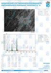
[15]E.V. Timchenko, Р.E. Timchenko, N.V. Tregub, L.A. Taskina, E.A. Selezneva Mapping of the Samara city bydefinition of areas with hydrogen degassing using Raman spectroscopy // Proc. of SPIE, 2014. - Vol. 9448. - No 94480K.
-
-
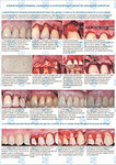
Хирургическое лечение множественных рецессий десны с использованием аллогенной твердой мозговой оболочки «Лиопласт» (Самара) (Лабораторное, Гистологическое, Клиническое, Рентгенологическое исследования)
 Презентация конференции
Хирургическое лечение множественных рецессий десны с использованием аллогенной твердой мозговой оболочки «Лиопласт» (Самара) (Лабораторное, Гистологическое, Клиническое, Рентгенологическое исследования)
Подробнее
Презентация конференции
Хирургическое лечение множественных рецессий десны с использованием аллогенной твердой мозговой оболочки «Лиопласт» (Самара) (Лабораторное, Гистологическое, Клиническое, Рентгенологическое исследования)
Подробнее
-
Клинико-экспериментальное обоснование нового метода хирургического лечения больных с ретенцией нижних третьих моляров
 Цель исследования — клинико-экспериментальное обоснование нового метода хирургического лечения больных с ретенцией нижних третьих моляров
Подробнее
Цель исследования — клинико-экспериментальное обоснование нового метода хирургического лечения больных с ретенцией нижних третьих моляров
Подробнее
-
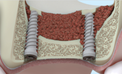
Оптимизация остеопластичес-кой коррекции атрофирован-ного альвеоляра челюсти. Клинико-экспериментальное исследование
 Цель исследования — разработка но-вого комплекса оперативной коррек-ции атрофированных альвеоляров челюстей для дентальной имплантации и протезирования
Подробнее
Цель исследования — разработка но-вого комплекса оперативной коррек-ции атрофированных альвеоляров челюстей для дентальной имплантации и протезирования
Подробнее
-
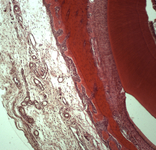
Морфологическое и лабора-торное обоснование приме-нения комбинированных трансплантатов при костной пластике челюстей
 Экспериментальное исследование. Изучение морфогенеза после заме-щения дефектов нижней челюсти смесью лиофилизированной алло-спонгиозы и “аллогенного гидроксиапатита” в соотношении 1:1 и 3:1.
Подробнее
Экспериментальное исследование. Изучение морфогенеза после заме-щения дефектов нижней челюсти смесью лиофилизированной алло-спонгиозы и “аллогенного гидроксиапатита” в соотношении 1:1 и 3:1.
Подробнее