+7 (929) 727 53 60 Травматология / Ортопедия
Analysis of the mineral component for cortical bone tissue by Raman spectroscopy after ovariectomy and its treatment with allogeneic hydroxyapatite
E. Timchenko1, P. Timchenko1, E. Pisareva1, M. Vlasov2, L. Volova2, I. Fedorova1, A. Tumchenkova1, M. Gorchenkova1 and A. Subatovich1
1 — Samara National Research University, Moskovskoye shosse 34, Samara, 443086, Russia
2 — Samara State Medical University, Chapayevskaya St. 89, Samara, 443099, Russia


In the work results of experimental researches of mineral component of cortical tissue after ovariectomy and estimation of correction using allogeneic hydroxyapatite (HAP) by the method of Raman spectroscopy are presented. A comparative analysis of bone samples from non-ovariectomized and ovariectomized animals after HAP injection was carried out using the Raman spectra. Coefficients which allow to estimate resorption of cortical tissue after ovariectomy and correction using HAP was entered. Introduction of HAP suspension showed that the consequences of ovariectomy can be almost compensated for the cortical tissue.
Lasers are widely used in the researches of musculoskeletal system diseases [1-4, 5]. Particular attention is drawn to the problem of frequent fractures of women due to the development of natural or artificial menopause which occurs after an early ovariectomy. The consequences of ovariectomy are detected as postovarioectomy syndrome and become the cause of osteoporosis, which is characterized by the development of destructive processes in mineral component of a bone, destruction of bone matrix and bone loss in general. Diagnosis of bone tissue damage is complicated by asymptomatic nature of the destructive processes, which lead to increased bone fragility and, as a consequence, to the osteopathic fractures. In the study of osteopathic changes in the bones after ovariectomy considerable attention is paid to the study of bone mineral density. Densitometry methods are widely used to determine bone mineral density. In clinical trials and medical practice the most widespread method is the method of dualenergy x-ray absorptiometry (DXA). However, this method can identify only 50% of osteopathic fractures of women [6]. Scanning electron microscopy (SEM) and transmission electron microscopy (TEM) are also used in the study of bone structure at the micro scale [7]. These general methods, which can be used for studying the hierarchical structure of bone at different levels of organization, are easy to apply, and most of them are non-destructive. However, they all focus on the structural and morphological area and cannot detect information about the composition. Spectroscopy methods [7-8] are the newest methods in the study of bio-tissues. The promising method is Raman spectroscopy [8]. It is easy to use, and also non-invasive and non-destructive method. The method provides simultaneous information on organic and inorganic components of samples with spatial resolution at the micro level. The aim of this work is to analyze the mineral component of cortical bone tissue by Raman spectroscopy after ovariectomy and its treatment with allogeneic hydroxyapatite.
Studies on modelling osteoporosis were conducted on female rats weighing up to 280 g [9]. All animals were divided into three groups: “ovariectomy” – a group of animals which was underwent bilateral ovariectomy, “non-operated with HAP” – a non-ovariectomized animals which had suspension HAP injected, and “ovariectomy with HAP” – a group of animals which had sterile suspension of allogenic gap in isotonic solution of sodium chloride injected into the thigh muscle. The bones were cut lengthwise and measured over the area of the entire section. Spectral characteristics of bones were obtained using the laboratory facility consisting of a diode laser (LML-785.0RB-04), an optical Raman probe (PBL 785), a spectrograph (Sharmrock SR-303i) with an integrated digital camera (ANDOR DV-420A-OE), and a computer. The application of the spectrograph is provided by a resolution of 0.15 nm (1 cm-1 ) and a low level of noise (reducing of the noise level, the matrix in the chamber is cooled to -60 °C [10]). A Raman probe focuses the laser radiation on the object (at a distance of 7.5 mm from the output window with a focal spot diameter of ~0.15 mm) and collects the backscattered radiation which is then sent by the optical fiber to the spectrometer. Spectra were recorded in the range from 400 cm-1 to 2000 cm-1 , as this spectral range is characterized by a relatively large depth of radiation penetration. The error in determining the using coefficients is estimated to be <4 % [11]. The spectrum processing was conducted using the software package Wolfram Mathematica 10. During the processing the researched spectrum was cleared up from noises using the smoothing median filter (5 points), the approximation line (a fifth order polynomial) of autofluorescent component was cm-1determined on the chosen interval of 400-2200 using an iteration algorithm, and then this component was subtracted and, as a result, the selected Raman spectrum was received [12].
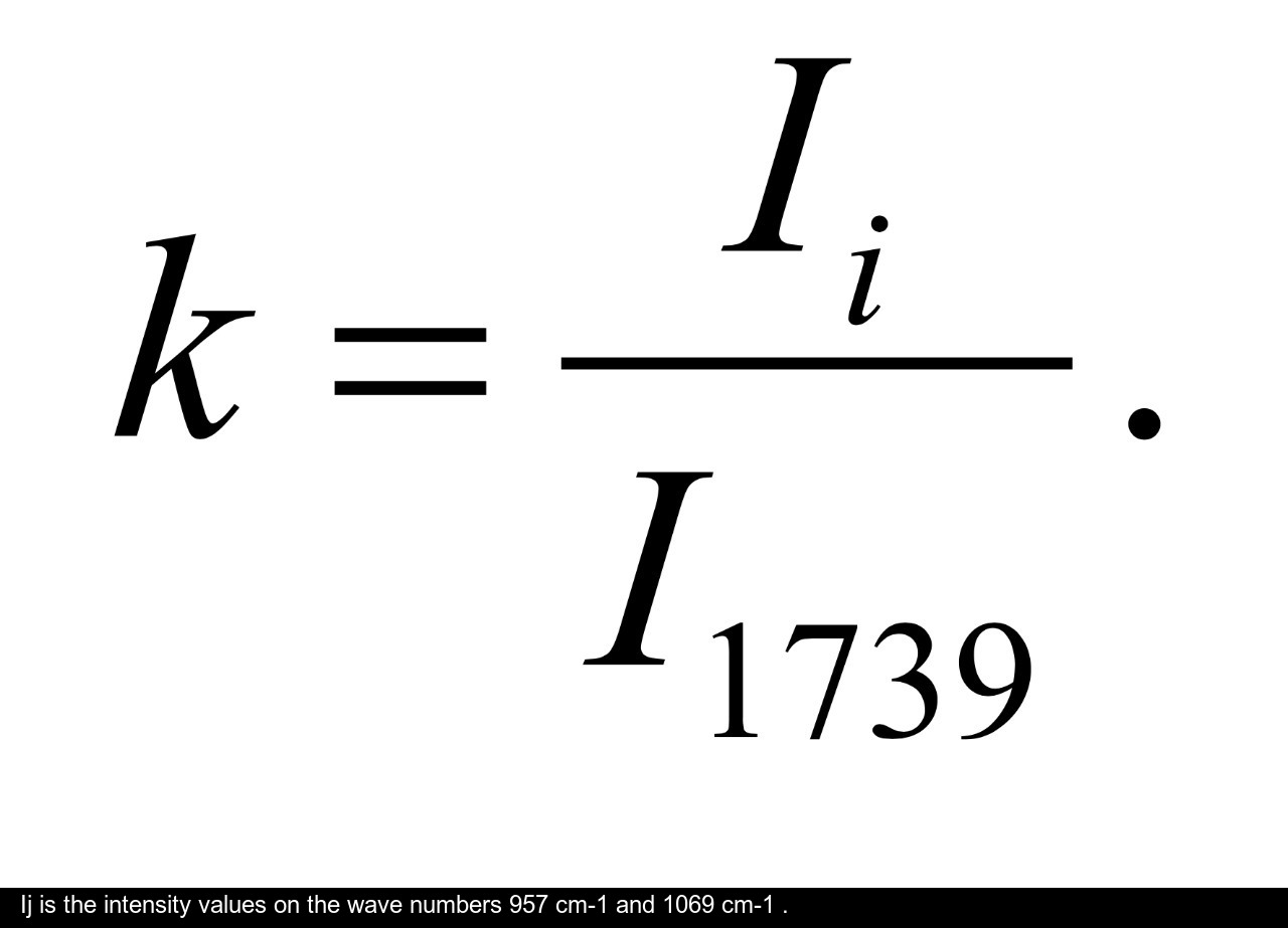

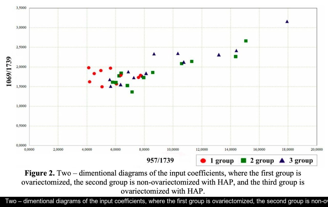
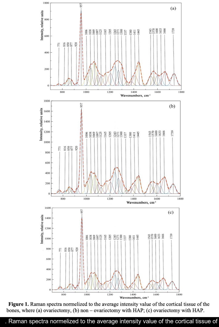
Figure 1 shows the averaged Raman spectra of cortical bone. Spectrum deconvolution was carried out in the software MagicPlotPro 2.7.2. The wavenumbers, that have main differences, are 957 cm-1 , 1038 cm-1 , 1069 cm-1 and 1243 cm-1 . Let us notice, that in osteoporosis the most significant changes occur in the mineral component of the bone at the wavenumbers 957 cm-1 ((v1) symmetric stretching vibration phosphate ion PO4 3- ) and 1069 cm-1 (C – O (v1) B – type substitution, planar valence oscillation) [13]. This is due to the substitutions of B – type phosphate ions PO4 3- on carbonates CO3 2- in the structure of hydroxyapatite. This process is a sign of resorption of cortical bone tissue, which leads to a destruction of the bone beams and their thinning. Figure 1 shows, that the ratio of peak intensity on the wave number 957 cm-1 (phosphate ion PO4 3- ) to peak intensity on the wave number 1069 cm-1 (CO3 2- ) in the samples with ovariectomy is less than in the samples with the introduced suspension HAP, indicating a partial restoration of bone beams. Optical coefficients were introduced to estimate the relative concentration of phosphate ions to carbonate ions in bone tissue of the samples:
Figure 2 shows that harmful consequences of ovariectomy identify the noted coefficients in decrease. The HAP treatment is showing that consequences of ovariectomy for cortical tissue of a bone can almost compensate. Perhaps, that is due to the features of cortical tissue location and structure (cortical bone tissue is the outer part of all skeletal structures).
Thus, major differences of intensity were found at the wave numbers 957 cm-1 and 1069 cm-1 for the samples of the rats with ovariectomy in comparison with the non-ovarectomized animals and samples with HAP. The entered coefficients allow to estimate the effectiveness of HAP to treat osteoresorption after modeling of osteoporosis using ovariectomy. It was identified that injection of HAP almost correct consequences of ovariectomy in cortical tissue.
The studies were carried out with financial support of Russian Foundation of Basic Research, project №18-315-20017\18.
-
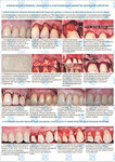
Research of component composition of bioimplants for treatment of gum recession using a Raman spectroscopy method
 Файл статьи P. Е. Timchenko1, Е. V. Timchenko1, L. Т. Volova2, О. О. Frolov1 and E. F. Yagofarova1Подробнее
 -
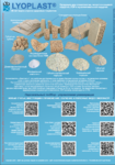
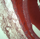
Raman spectroscopy method for the evaluation of bone bioimplants made using the "Lyoplast" technology from cadaveric and in vivo resected bone tissue
 Файл статьи P. Е Timchenko1, Е. V Timchenko1, L. Т Volova2, D. А Dolgushkin , V. V Boltovskaya , О. О Frolov1Подробнее
 -
Raman spectroscopy for evaluation of dura mater based graft
 Файл статьи P. Е Timchenko, Е. V Timchenko, L. Т Volova, О. О Frolov, А. U Kulabuhova, N. K KiykoПодробнее
 -
Хирургическое лечение множественных рецессий десны с комбинированным применением аутотрансплантата и аллогенной лиофилизированной dura mater: клинический случай
 Файл статьи
М. А. Носова1, Л. Т. Волова1, А. Н. Шаров2, Д.А. Трунин1, М. А. Постников1
Подробнее
Файл статьи
М. А. Носова1, Л. Т. Волова1, А. Н. Шаров2, Д.А. Трунин1, М. А. Постников1
Подробнее