+7 (929) 727 53 60 Травматология / Ортопедия
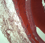
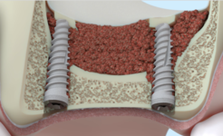
Determination of histological composition at the locus of installation of allogeneic dura mater implant an in vivo model: laborator-histomorphological research
М. А. Носова, Л. Т. Волова, А. Н. Шаров, Д.А. Трунин, И.Ф. Нефедова
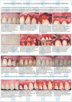
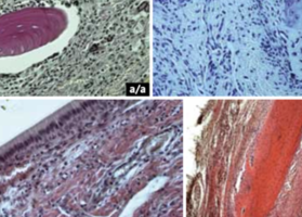
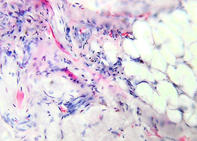
Objective. Gingival recession is a common pathology in Russia and worldwide. The modern techniques of the surgical treatment allow completely eliminating the signs of recession with the adequate choice of the surgical treatment strategy, tactics, technique and surgical protocol, provide the absence of complications and stable results in the long term. The use of autograft and allogeneic dura mater as a plastic material to create/increase the volume of the attached gingiva in the treatment of gingival recession have comparable results in all clinical parameters. The reason for the stability of the result and the absence of recurrence is poorly studied; the data are fragmentary and do not give an understanding of the full picture. Analysis of the histological composition of tissues in the area of plastic material placement is not found in the scientific literature. In case of recession the plastic material is placed partially subperiosteal (in the area of the tooth root), partially into the thickness of the soft tissues of the gum. The aim of the work is to determine the histological composition of the tissues at the dura mater implant site compared to the control without it; to assess the changes in the implanted structure and tissue reaction of the surrounding tissues as a result of the operation at the cellular level. Materials and methods. A laboratory histomorphological study was carried out on 60 laboratory rats. All of them were subjected to the surgery adequate to the surgical treatment technique of the gingival recession: in the control group without plastic material, in the study group – with allogenic dura mater. Macro preparations were taken on the 3-7-14-28-90-107 days after the surgery. Results. In all cases the tissue complex is repeatedly formed and the reaction to the operation is the same. The replacement of the plastic material occurs in the same terms. At the subperiosteal instillation the plastic material is replaced by bone tissue, at the intragingival - by connective tissue. The thickening of the gingival biotype occurs to a large extent to a large extent due to trauma from the surgery, less from the plastic material. Allogeneic dura mater stimulates ossification earlier in comparison with the control. Conclusions. In all cases of plastic material application in surgical treatment of recessive It is justified to install it subperiostally, forming the full-layer muco-periosteal flap using an acute method (scalpel) to preserve the cambial layer of the periosteum. Restoring/creating the bone volume of the alveolar lamina closure vestibularly provides support to the soft tissues of the gingiva; determines the stability of the result of surgical gingival recession treatment and a good prognosis in the long term: without complications and recurrence.
-
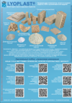
Постимплантационный гистогенез в месте применения аллогенной dura mater в лабораторном эксперименте на крысах. Лабораторно-гистоморфологическое исследование
 Файл статьи
Подробнее
Файл статьи
Подробнее
-
Научно-практический журнал «Академия Хлорофилла и коры Осины» Том 2, Выпуск 1-2(3-4) март 2024.
 Подробнее
-
Научно-практический журнал Академия Хлорофилла и Коры Осины Том №1, Выпуск 2 (2) Декабрь 2023
 Подробнее
-
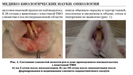
Клинико-экспериментальное обоснование нового метода хирургического лечения больных с ретенцией нижних третьих моляров
 Цель исследования — клинико-экспериментальное обоснование нового метода хирургического лечения больных с ретенцией нижних третьих моляров
Подробнее
Цель исследования — клинико-экспериментальное обоснование нового метода хирургического лечения больных с ретенцией нижних третьих моляров
Подробнее